
Quick answer: Subcutaneous fat lies beneath the skin, while visceral fat sits deeper in the abdomen around internal organs. Their locations and associations with health risk differ. A pinch, belly shape, or photo cannot measure visceral fat. Waist measurements support risk screening; medical assessment and imaging provide information that external appearance cannot.
You can pinch a fold at your waist, yet your scale gives you a separate "visceral fat" rating. You now have 2 observations and no clear way to tell whether they describe the same tissue. They do not measure the same thing.
Visceral fat vs subcutaneous fat is primarily a distinction about location. Understanding that distinction helps you interpret measurements without treating a belly shape, a scale score, or a progress photo as a diagnosis.
What is the difference between visceral fat and subcutaneous fat?
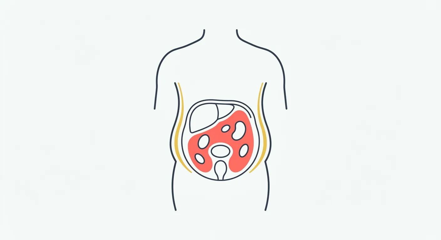
Subcutaneous fat lies under your skin. Visceral fat lies deeper in your abdomen around internal organs. Reports may abbreviate visceral adipose tissue as VAT. Both are normal parts of the body; the amount, distribution, and wider clinical picture matter when considering health risk.
| Question | Subcutaneous fat | Visceral fat |
|---|---|---|
| Where is it stored? | Beneath the skin, including the abdomen, hips, thighs, and arms | Inside the abdomen around internal organs |
| Does a pinch reach it? | A skinfold includes skin and underlying subcutaneous tissue | No; a pinch cannot reach the internal compartment |
| Can a photo measure it? | No; the visible outline includes several tissues and is affected by viewing conditions | No; a photograph cannot see inside the abdomen |
| What is its role? | Includes energy storage, insulation, cushioning, and metabolic functions | An internal fat depot with metabolic functions and health-risk associations |
| What does its amount mean for health? | Interpretation depends on amount, distribution, and other findings | Greater visceral adiposity is associated with cardiometabolic risk, but appearance cannot diagnose it |
| How can compartments be assessed? | Anatomical imaging can distinguish the outer fat compartment | CT/MRI can depict it; compatible DXA software can estimate VAT |
Why does fat location matter for health?
Subcutaneous tissue is more than a cosmetic layer. It stores energy, helps insulate and cushion the body, and participates in metabolic signaling. You cannot classify all outer fat as harmless or all internal fat as abnormal.
Central adiposity is associated with health risks, including type 2 diabetes and cardiovascular disease. NIDDK explains that a larger waist can indicate increased risk even at a normal BMI. Your family history, activity, medical history, and other findings also matter.
Fat distribution varies between people. A lean-looking waist cannot guarantee low internal fat, and a visible belly cannot establish how much fat surrounds the organs. A clinician can interpret your risk without relying on appearance alone.
Can you tell which type you carry by pinching or feeling your belly?
A pinch reaches skin and subcutaneous tissue. It cannot tell you how much fat surrounds your organs. Belly firmness and protrusion have multiple causes and should not be used to diagnose visceral fat.
Muscle tension and abdominal distension can affect how your belly feels. A larger or smaller skinfold does not establish which compartment accounts for most abdominal fat. There is no reliable hard-belly versus soft-belly test for that distinction.
Our explanation of why appearance cannot identify visceral fat covers the limits of belly shape, firmness, and photo comparisons. Combining those observations does not produce an internal fat measurement.
Which measurements can distinguish the fat compartments?
Some methods estimate overall body composition; others assess a particular region or tissue compartment. Read the output name and unit before comparing results.
| Method | What the result represents | Interpretation limit |
|---|---|---|
| Waist circumference | External abdominal circumference used in risk screening | Includes more than visceral fat and cannot separate compartments |
| Skinfold measurement | A fold of skin and underlying subcutaneous tissue at a selected site | Cannot measure visceral fat or all subcutaneous fat directly |
| DXA android fat | Fat estimated in a defined abdominal region | Not interchangeable with the region's visceral fat estimate |
| DXA VAT estimate | A modeled estimate generated by compatible scanner software | Method-specific, with measurement error |
| CT or MRI | Images that distinguish anatomical compartments | Requires appropriate acquisition, analysis, and clinical interpretation |
| Progress photo | External appearance | Cannot isolate or quantify either fat compartment |
NICE recommends waist-to-height ratio alongside BMI for central-adiposity and health-risk assessment in adults with BMI below 35. A shrinking waist is a change worth recording. It does not establish how much of that change came from visceral fat.
A comparison of iDXA and MRI illustrates why method details matter: the VAT results were strongly related, but the study also found bias and differences between individual measurements. Ask whether a DXA report contains a dedicated VAT estimate or only total android fat.
A clinician can decide whether CT, MRI, or another assessment would answer a relevant question for you. CT involves radiation, and neither a home photo nor curiosity about a belly shape creates a universal need for imaging. Our overview of body fat measurement methods explains how different outputs fit together.
Do nutrition and activity changes affect both compartments?
Research suggests that nutrition and activity changes can affect abdominal fat, with responses that vary between people and interventions. A randomized diet-and-exercise study assessed changes using MRI; a separate exercise trial examined individual responses. Neither gives you a personal countdown for when each depot will change.
You cannot infer that unchanged photos mean visceral fat is being lost first, or that a flatter belly confirms a particular internal change. Keep nutrition and activity decisions appropriate to your health and goals, and use clinical advice when you need individual interpretation.
What can progress photos tell you over time?
Photos record external appearance under the conditions used. Lighting, posture and other factors can change how a body looks; a photo comparison cannot isolate or quantify either fat depot. You can compare external appearance over time while keeping those limits attached to the result.

A comparison documents appearance. It does not identify which tissue caused a visible change.
GainFrame provides an iOS photo library and AI estimates from selected progress images. A falling estimate cannot confirm a calorie deficit, isolate subcutaneous fat loss, or establish visceral fat reduction.
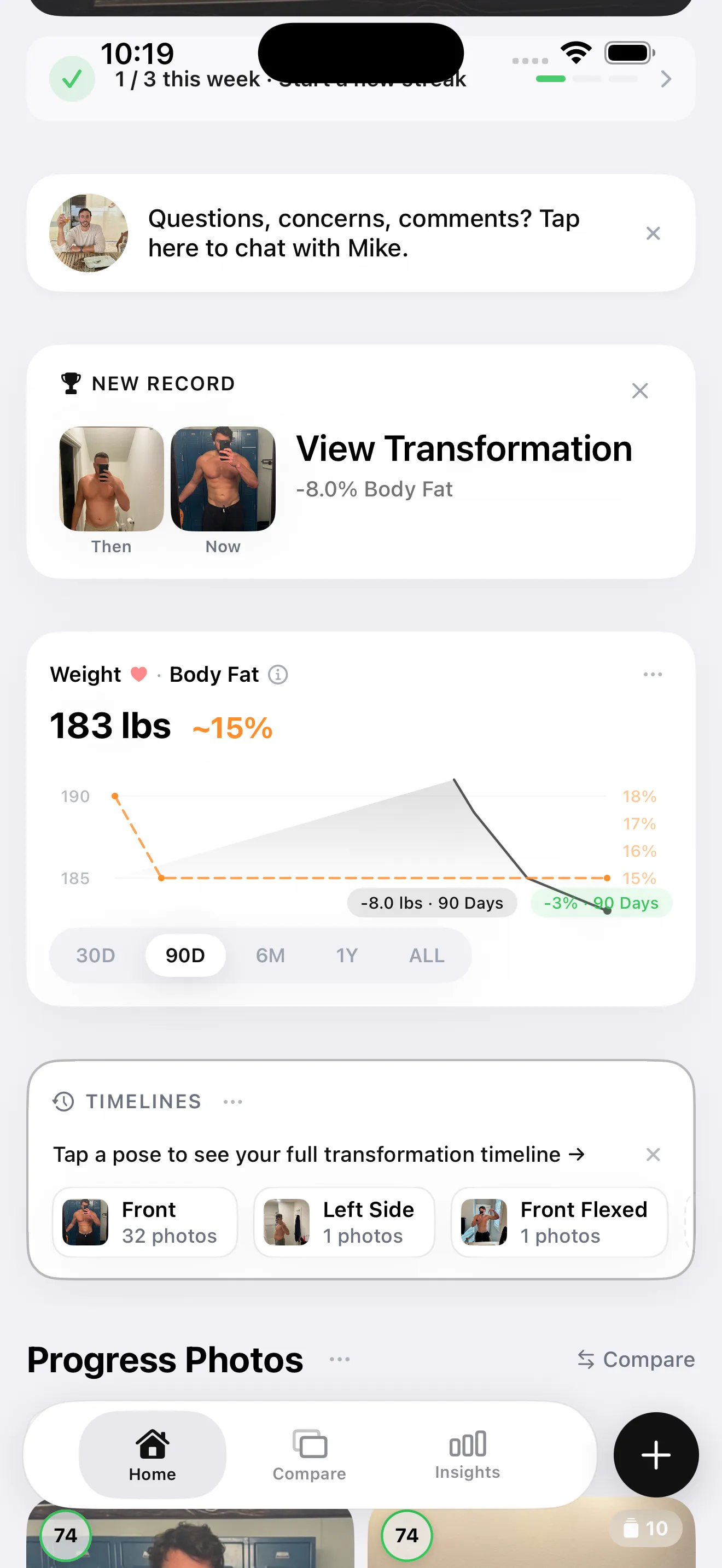
Keep the method beside the number. A photo-derived estimate is different from an imaging assessment.
How should you keep track of the different measurements?
Choose records that match the question you want to answer:
- For visible changes: keep camera height, distance, lighting, and posture consistent across photos.
- For waist changes: use a repeatable tape-measure protocol and record the measurement site.
- For a device score: keep the manufacturer, model, unit, and conditions with the reading.
- For health questions: bring relevant records to a clinician, who can assess them alongside your history and other findings.
A useful record preserves these distinctions. You do not need to label every visual change as visceral or subcutaneous to document it accurately.
What else should you know about visceral fat?
Where is each type of fat stored?
Subcutaneous fat sits under the skin across areas such as the abdomen, hips, thighs, and arms. Visceral fat sits deeper in the abdomen around internal organs. Both are normal anatomical compartments. Their location matters, but your appearance cannot show how much of your abdominal fat belongs to each compartment.
Can you tell which type you have by pinching?
Pinching gathers skin and underlying subcutaneous tissue. It does not reach visceral fat or establish which compartment accounts for most of your abdominal fat. A large or small skinfold cannot rule out an internal fat concern. Clinical assessment and suitable measurement methods answer questions that a pinch cannot resolve.
Does a hard belly mean visceral fat?
Firmness is not a reliable test for visceral fat. Muscle tension, abdominal distension, and other factors can change how your belly feels, and a soft area does not rule out internal fat. You cannot diagnose a fat distribution pattern by pressing on your abdomen. Ask a clinician about concerns or persistent changes.
Can a DEXA scan distinguish the compartments?
Some DXA systems use compatible software to estimate visceral adipose tissue within a defined abdominal region. That output differs from total android fat and depends on the equipment and analysis method. CT and MRI can depict the anatomical compartments. A clinician can help interpret the report and decide whether further imaging is appropriate.
Can progress photos track visceral fat?
Photos cannot see or measure visceral fat. They also cannot isolate the amount of subcutaneous fat from the other tissues affecting your appearance. Consistent images provide a useful visual record, but lighting, posture, and other factors influence them. Neither visible change nor an unchanged photo establishes what happened to internal fat.